Integumentary System Quiz
88 multiple-choice questions with answers and explanations
Test yourself on the skin and its accessory structures: the epidermal layers and cells, the dermis and hypodermis, skin color, hair and nails, sweat and sebaceous glands, how the skin protects, senses and cools the body, skin cancers and common disorders, and how skin heals.
Read a question, pick an option, then open the answer to check yourself and read why it is correct.
Epidermal layers and cells
Question 1
Which tissue type forms the epidermis?
Show answer
Answer: A. Keratinized stratified squamous epithelium
The epidermis has multiple layers of flattened surface cells that become keratinized. This arrangement suits its protective role.
Question 2
Which statement describes the blood supply within the epidermis?
Show answer
Answer: B. It contains no blood vessels.
The epidermis is avascular. Blood vessels are present in the deeper connective tissues.
Question 3
Which sequence lists the epidermal strata of thin skin from deepest to most superficial?
Show answer
Answer: C. Basale, spinosum, granulosum, corneum
Thin skin has four epidermal strata. New cells originate in the basal layer and move toward the surface.
Question 4
A sample from a palm has an epidermal layer between the stratum granulosum and stratum corneum. Which layer is it?
Show answer
Answer: D. Stratum lucidum
The stratum lucidum is the extra epidermal stratum in thick skin. It lies between the granular and cornified strata.
Question 5
Which intracellular fibrous protein gives skin, hair, and nails their hardness and water-resistant properties?
Show answer
Answer: A. Keratin
Keratin is a fibrous protein stored inside keratinocytes. Collagen and elastin instead support the connective tissues of the skin.
Question 6
Which epidermal layer contains the stem cells that continually produce new keratinocytes?
Show answer
Answer: B. Stratum basale
Basal stem cells divide in the deepest epidermal stratum. Their descendants replace cells shed at the surface.
Question 7
What happens to existing epidermal cells as new cells form in the stratum basale?
Show answer
Answer: C. They are pushed toward the skin surface.
Cell production in the basal layer displaces older cells superficially. Those cells undergo changes as they move through the epidermal strata.
Question 8
Which cells in the stratum basale stimulate sensory nerves involved in touch?
Show answer
Answer: D. Merkel cells
Merkel cells serve a sensory role in the basal layer. Melanocytes in that same layer produce pigment instead.
Question 9
Which junctions are associated with the spiny appearance of stained stratum spinosum?
Show answer
Answer: A. Desmosomes
Desmosomes interlock to reinforce attachment between cells. They contribute to the characteristic appearance of stained stratum spinosum.
Question 10
Which epidermal cells engulf bacteria, foreign particles, and damaged cells in the stratum spinosum?
Show answer
Answer: B. Langerhans cells
Langerhans cells are dendritic immune cells that act as phagocytes in this layer. Their role differs from pigment production and touch detection.
Question 11
What happens to nuclei and other organelles as keratinocytes die in the stratum granulosum?
Show answer
Answer: C. They disintegrate.
The loss of nuclei and organelles accompanies keratinocyte death. Proteins and cell membranes remain to contribute to the superficial barrier.
Question 12
Which clear protein gives the stratum lucidum its translucent appearance?
Show answer
Answer: D. Eleidin
Eleidin is derived from keratohyalin and fills the dead, flattened cells of the stratum lucidum. Its clarity gives this layer its name.
Question 13
Which description best fits the keratinocytes of the stratum corneum?
Show answer
Answer: A. Dead cells that are regularly shed
The surface stratum consists of dead keratinized cells that slough off. Cells from deeper strata continually replace them.
Dermis, hypodermis and skin color
Question 14
Which connective tissue forms the papillary layer of the dermis?
Show answer
Answer: B. Loose areolar connective tissue
The papillary dermis has a loose mesh of collagen and elastin. The deeper reticular layer is dense irregular connective tissue.
Question 15
Which dermal layer is thicker and made of dense irregular connective tissue?
Show answer
Answer: C. Reticular layer
The reticular layer lies deep to the papillary layer. Its dense connective tissue provides substantial structural support.
Question 16
Which statement correctly relates dermal papilla folding to attachment between the epidermis and dermis?
Show answer
Answer: A. More folding produces stronger attachment.
Dermal papillae increase the strength of attachment between the two layers. Greater folding strengthens their connection.
Question 17
Which dermal fibers allow the skin to stretch and recoil during movement?
Show answer
Answer: A. Elastin fibers
Elastin supplies elasticity. Collagen primarily supplies structure and tensile strength.
Question 18
Which layer connects the skin to underlying fascia but is not strictly part of the skin?
Show answer
Answer: C. Hypodermis
The hypodermis lies beneath the dermis and attaches skin to deeper structures. OpenStax distinguishes it from the skin itself.
Question 19
Which function of hypodermal adipose tissue directly protects deeper structures against mechanical trauma?
Show answer
Answer: C. Mechanical cushioning
Hypodermal fat cushions underlying structures against trauma; it also stores energy and provides thermal insulation.
Question 20
Which vesicles transfer pigment from melanocytes into keratinocytes?
Show answer
Answer: A. Melanosomes
Melanosomes carry melanin into keratinocytes. This distributes pigment among epidermal cells.
Question 21
Which comparison of the two main forms of melanin is correct?
Show answer
Answer: B. Eumelanin is black or brown; pheomelanin is red.
OpenStax distinguishes black and brown eumelanin from red pheomelanin. Both are forms of melanin.
Question 22
How does increased melanin accumulation after UV exposure help epidermal cells?
Show answer
Answer: C. It protects their DNA from UV damage.
The pigment accumulated in keratinocytes offers protection against UV injury. This protection does not imply that all UV damage is prevented.
Question 23
What defect primarily accounts for the reduced pigmentation in albinism?
Show answer
Answer: D. Melanocytes cannot produce normal amounts of melanin.
Albinism is a genetic disorder involving impaired melanin production. It can affect pigmentation of skin, hair, and eyes.
Question 24
Which pigment disorder causes patches of skin to lose color when local melanocytes lose their ability to produce melanin?
Show answer
Answer: A. Vitiligo
Vitiligo produces localized patches of depigmentation. Albinism is a genetic pigmentation disorder rather than this pattern of localized loss.
Question 25
A prolonged reduction in blood oxygen makes the skin appear blue. What is this color change called?
Show answer
Answer: B. Cyanosis
Cyanosis reflects an increased influence of deoxygenated hemoglobin on skin color. It does not result from a change in melanin production.
Hair structure and growth
Question 26
From which skin layer do accessory structures such as hair follicles originate during development?
Show answer
Answer: C. Epidermis
Accessory structures begin as epidermal structures. They can extend into the deeper dermis and hypodermis.
Question 27
Which description distinguishes the hair root from the hair shaft?
Show answer
Answer: D. The root is anchored in the follicle below the skin surface.
The hair root is the portion retained within the follicle beneath the surface. The shaft is the portion not anchored to the follicle.
Question 28
Which structure in the hair bulb contains the mitotically active basal cells that produce hair?
Show answer
Answer: A. Hair matrix
The hair matrix contains dividing cells. Their descendants are pushed outward and become keratinized hair.
Question 29
Which structure enclosed by the hair bulb contains connective tissue, capillaries, and nerve endings?
Show answer
Answer: B. Hair papilla
The papilla brings dermal connective tissue and its vessels and nerves into the bulb. It is distinct from the keratinized layers of the hair.
Question 30
Which layer forms the central core of a hair when that layer is present?
Show answer
Answer: C. Medulla
The medulla is the central core of a hair. Some hairs lack this layer.
Question 31
Which hair layer forms the outer covering of very hard, keratinized cells?
Show answer
Answer: D. Cuticle
The cuticle is the outer covering of the hair itself. It surrounds the cortex rather than forming the central core.
Question 32
Why does cutting the externally visible hair shaft not produce sensation in the hair itself?
Show answer
Answer: A. The external hair is completely dead and keratinized.
The exposed hair shaft is dead and keratinized. Sensory nerves associated with the follicle can detect movement, but cutting the exposed shaft does not stimulate sensation in the hair itself.
Question 33
Which follicle structure is a basement membrane between the external root sheath and its connective tissue sheath?
Show answer
Answer: B. Glassy membrane
The glassy membrane separates the epithelial external root sheath from the surrounding connective tissue. It is not a layer of the hair shaft.
Question 34
Which protective role does OpenStax specifically attribute to eyelashes and hairs in the nose and ears?
Show answer
Answer: C. Trapping dust particles that may carry allergens or microbes
These hairs help prevent entry of unwanted particles. Their protective role depends on trapping and excluding material.
Question 35
Which muscle contracts to make a hair stand up during goose bumps?
Show answer
Answer: D. Arrector pili
The arrector pili is smooth muscle connected to a hair root. Sympathetic stimulation causes its contraction and raises the shaft.
Question 36
Which statement correctly describes the active growth phase of the hair cycle?
Show answer
Answer: A. Anagen involves rapid cell division at the root and outward shaft growth.
Anagen is the active growth phase, when cells divide rapidly at the hair root and push the shaft outward. Catagen is transitional, and telogen is the resting phase.
Question 37
Which statement correctly identifies the resting phase of the hair cycle?
Show answer
Answer: B. Telogen is the resting phase, with no new growth.
Telogen is the resting phase, lasting about 2 to 4 months; catagen is a transition lasting 2 to 3 weeks, and anagen is active growth.
Question 38
Which change contributes to hair becoming gray or white with age?
Show answer
Answer: C. Decreased melanin production
As pigment production declines, hair loses its original color. This is a change in pigmentation rather than sweat or oil secretion.
Nails and skin glands
Question 39
What cells make up the nail body?
Show answer
Answer: D. Densely packed dead keratinocytes
The nail body is composed of dead keratinized cells. Its dense structure helps protect the ends of the digits.
Question 40
Where do proliferating basal cells allow a nail to grow continuously?
Show answer
Answer: A. In the matrix at the nail root
The nail root contains a matrix of cells derived from the stratum basale. Their proliferation supports continued nail growth.
Question 41
Which structure overlaps the nail at its sides and helps anchor the nail body?
Show answer
Answer: B. Lateral nail fold
The lateral folds secure the sides of the nail. The lunula is a pale crescent over the matrix rather than a side fold.
Question 42
Which nail structure is also called the eponychium?
Show answer
Answer: C. Nail cuticle
The eponychium is the cuticle at the proximal end of the nail body. It is formed by the proximal nail fold.
Question 43
What is the crescent-shaped region at the base of the nail called?
Show answer
Answer: D. Lunula
The lunula forms where thick epithelium overlies the nail matrix. The rest of the vascular nail bed generally appears pink.
Question 44
Which region beneath the free edge of a nail consists of thickened stratum corneum?
Show answer
Answer: A. Hyponychium
The hyponychium lies under the distal free edge. The eponychium is the proximal cuticle.
Question 45
By which mechanism do sweat gland cells release their secretions?
Show answer
Answer: B. Exocytosis
OpenStax classifies sweat glands as merocrine glands, which release secretions by exocytosis without loss of the secretory cell.
Question 46
Which gland produces hypotonic sweat as a major mechanism of thermoregulation?
Show answer
Answer: C. Eccrine sweat gland
Eccrine sweat is mainly water with dissolved substances. Its release supports temperature regulation.
Question 47
Where does an apocrine sweat gland duct normally release its secretion?
Show answer
Answer: B. Into a hair follicle
Apocrine glands are usually associated with hair follicles in densely hairy areas. Their ducts normally release sweat into the follicle.
Question 48
Why is apocrine sweat particularly associated with body odor?
Show answer
Answer: A. Its organic compounds undergo bacterial decomposition.
Apocrine sweat contains organic material in addition to water and salts. Bacterial breakdown of that material produces odor.
Question 49
How do aluminum-based antiperspirants reduce the sweat reaching the skin surface?
Show answer
Answer: B. They form a physical block in sweat gland ducts.
The aluminum compounds precipitate inside the duct as its pH changes. The resulting obstruction limits sweat release.
Question 50
Which product of sebaceous glands lubricates the stratum corneum and helps keep it pliable?
Show answer
Answer: C. Sebum
Sebum is a lipid mixture that lubricates the dry, keratinized surface. It also helps reduce water loss.
Protection, sensation and temperature
Question 51
Which combination in the stratum corneum helps form the barrier against water loss?
Show answer
Answer: A. Keratin and glycolipids
Keratin and glycolipids contribute to the surface barrier that limits water loss. Collagen and elastin primarily support dermal connective tissue.
Question 52
Which substance in sweat helps deter microbial overgrowth on the skin?
Show answer
Answer: B. Dermcidin
Dermcidin has antimicrobial properties. This gives sweat a protective role in addition to cooling.
Question 53
Which encapsulated cutaneous receptor responds to light touch?
Show answer
Answer: C. Meissner corpuscle
Meissner corpuscles are tactile receptors for light touch. Pacinian corpuscles respond to vibration.
Question 54
Which receptor, located deep in the dermis and hypodermis, responds to deep pressure and vibration?
Show answer
Answer: D. Pacinian corpuscle
Pacinian (lamellated) corpuscles detect deep pressure and high-frequency vibration around 250 Hz, whereas Meissner (tactile) corpuscles detect light touch and vibrations below 50 Hz.
Question 55
Which structure first detects a disturbance of a hair and sends sensory information toward the central nervous system?
Show answer
Answer: A. Hair root plexus
The nerve plexus surrounding a follicle detects movement of the hair. It transmits information to the brain and spinal cord.
Question 56
At what step does sweat directly remove heat from the skin?
Show answer
Answer: B. When it evaporates from the surface
Evaporation dissipates body heat into the environment. Simply producing sweat is not the cooling step described here.
Question 57
Why can the skin become red during vigorous exercise?
Show answer
Answer: B. Dermal arterioles dilate, increasing blood flow near the surface.
Dilation increases the delivery of warm blood to the skin. This promotes heat dissipation and can cause visible redness.
Question 58
Which change in dermal arterioles and surface blood flow helps conserve body heat in a cold environment?
Show answer
Answer: D. Arteriolar constriction with decreased surface blood flow
Arteriolar constriction reduces heat delivery to the surface. This helps keep internal structures warm.
Question 59
Which division of the nervous system helps coordinate skin responses for temperature regulation?
Show answer
Answer: A. Sympathetic nervous system
OpenStax links integumentary temperature regulation to the sympathetic division. It helps initiate sweating and other temperature-related responses.
Question 60
How does mitotic activity in the stratum basale change with aging in a way that contributes to epidermal thinning?
Show answer
Answer: B. It decreases.
Reduced basal cell division slows the supply of new epidermal cells. This contributes to thinning of the epidermis.
Question 61
Which substance is the precursor for cholecalciferol synthesis in sun-exposed skin?
Show answer
Answer: D. A derivative of cholesterol
UV exposure enables synthesis of cholecalciferol from a cholesterol derivative. This begins the pathway that produces active vitamin D.
Question 62
After cholecalciferol is formed in skin, which sequence leads to active calcitriol?
Show answer
Answer: A. Liver forms calcidiol, then kidneys form calcitriol.
Vitamin D activation involves both liver and kidneys. The liver makes calcidiol, and the kidneys then produce active calcitriol.
Skin cancers and common disorders
Question 63
How can excessive UV exposure contribute to skin cancer?
Show answer
Answer: B. By damaging DNA and allowing mutations to accumulate
UV injury can damage cellular DNA. Accumulated mutations can permit abnormal survival and uncontrolled proliferation.
Question 64
What is the term for cancer cells spreading and establishing tumors in other organs?
Show answer
Answer: C. Metastasis
Metastasis is the spread of cancer cells from the original site to establish tumors in other organs.
Question 65
Which cells are affected in basal cell carcinoma?
Show answer
Answer: D. Mitotically active stem cells of the stratum basale
Basal cell carcinoma arises in the epidermal basal stem-cell population. Squamous cell carcinoma affects keratinocytes of the stratum spinosum.
Question 66
Which skin cancer affects keratinocytes of the stratum spinosum?
Show answer
Answer: A. Squamous cell carcinoma
Squamous cell carcinoma involves spinous-layer keratinocytes. Its cell of origin differs from that of basal cell carcinoma and melanoma.
Question 67
Which cell population grows uncontrollably in melanoma?
Show answer
Answer: B. Melanocytes
Melanoma is a cancer of pigment-producing melanocytes. OpenStax describes it as highly metastatic.
Question 68
Which characteristic most directly accounts for the high fatality of melanoma?
Show answer
Answer: C. Its high metastatic potential before detection
Melanoma can spread to other organs before it is recognized. That metastatic behavior contributes to its high fatality.
Question 69
How does OpenStax compare the aggressiveness of squamous cell carcinoma with basal cell carcinoma?
Show answer
Answer: C. Squamous cell carcinoma is more aggressive.
OpenStax describes squamous cell carcinoma as more aggressive than basal cell carcinoma. Untreated squamous cell carcinoma can metastasize.
Question 70
In the ABCDE warning signs for melanoma, what does evolving mean?
Show answer
Answer: A. The shape of the mole has changed.
Evolving refers to a change in the lesion. A changing mole is a reason to seek clinical assessment rather than diagnose it from appearance alone.
Question 71
Which disorder is described as producing dry, itchy patches that resemble rashes?
Show answer
Answer: B. Eczema
OpenStax describes eczema as an inflammatory condition with dry, itchy skin patches. Acne instead involves blocked follicles and associated inflammation.
Question 72
Which treatments does OpenStax list for managing eczema symptoms?
Show answer
Answer: C. Moisturizers, corticosteroid creams, and immunosuppressants
OpenStax lists moisturizers, corticosteroid creams, and immunosuppressants for managing eczema symptoms.
Question 73
Which accumulation can plug a hair follicle during acne development?
Show answer
Answer: D. Excess sebum together with keratin
Excess sebum mixes with keratin to obstruct the follicle. The resulting plug can contribute to acne lesions.
Question 74
What makes an initially white sebum plug turn black when exposed to air?
Show answer
Answer: A. Oxidation of the sebum
The color change comes from oxidation after air exposure. OpenStax does not attribute the black color to dirt.
Question 75
Which secretion is stimulated by androgens and contributes to follicular plugging during puberty-associated acne?
Show answer
Answer: B. Sebum
Androgens stimulate sebum release. Excess sebum together with keratin can block hair follicles and contribute to acne.
Injuries and skin repair
Question 76
A cut damages dermal blood vessels and causes bleeding. Which early event in skin repair helps stop the blood loss?
Show answer
Answer: C. Formation of a blood clot
A blood clot helps stop bleeding from damaged vessels and later dries into a scab; fibroblasts subsequently deposit collagen as tissue repair proceeds.
Question 77
Which cells deposit collagen to form granulation tissue during skin repair?
Show answer
Answer: D. Fibroblasts
Fibroblasts multiply and lay down collagen in the wound. Capillary growth then helps improve oxygen delivery to the repair area.
Question 78
How does extensive destruction of the protective skin barrier affect fluid loss from the body?
Show answer
Answer: A. It increases fluid loss.
Destruction of the skin barrier allows major fluid loss. This can lead to electrolyte imbalance and circulatory and renal failure.
Question 79
Loss of which skin function most directly explains the increased infection risk after a severe burn?
Show answer
Answer: B. Physical protection against pathogen entry
Burns breach the physical barrier that normally helps exclude pathogens. The exposed tissues are therefore particularly vulnerable.
Question 80
A mild sunburn damages only the epidermis. How is it classified?
Show answer
Answer: C. First-degree burn
First-degree burns are superficial and limited to the epidermis. Deeper burn categories involve the dermis or underlying tissues.
Question 81
Which burn category damages the epidermis and part of the dermis and commonly causes painful blistering?
Show answer
Answer: D. Second-degree burn
Second-degree burns extend into a portion of the dermis. OpenStax associates this depth with swelling and painful blisters.
Question 82
A burn destroys the entire epidermis and dermis and damages sensory nerve endings, but underlying muscle and bone are not involved. Which category best matches this description?
Show answer
Answer: A. Third-degree burn
Third-degree burns destroy the full thickness of the skin. Fourth-degree burns extend farther into muscle and bone.
Question 83
A burn extends through the skin into underlying muscle and bone. How is it classified?
Show answer
Answer: B. Fourth-degree burn
Fourth-degree burns involve tissues beneath the skin, including muscle and bone. This makes them more extensive in depth than third-degree burns.
Question 84
Why can third- and fourth-degree burns be less painful than shallower burns?
Show answer
Answer: C. Damage to the sensory nerve endings themselves
Deep burns can destroy the structures that would otherwise transmit pain. Reduced pain therefore does not indicate a minor injury.
Question 85
How does mature scar tissue differ from regenerated normal skin?
Show answer
Answer: D. It is collagen-rich and does not regenerate normal accessory structures.
Scar repair deposits collagen without recreating the original skin architecture. Hair follicles and skin glands do not regenerate within this fibrous repair tissue.
Question 86
Rapid expansion of the body stretches the dermis beyond its elasticity. Which skin change can result?
Show answer
Answer: A. Stretch mark
Stretch marks develop when stretching exceeds the elastic limits of the dermis. They can accompany rapid weight gain during puberty or pregnancy.
Question 87
Which sequence explains bedsore formation after prolonged pressure over a bony area?
Show answer
Answer: B. Pressure reduces local blood flow, leading to tissue death.
Sustained pressure compromises local circulation and can cause necrosis. Repositioning helps relieve the pressure responsible.
Question 88
Which cellular response does OpenStax describe as causing a callus after repeated abrasion from a poorly fitting shoe?
Show answer
Answer: C. Increased division of basal epidermal stem cells
Repeated friction stimulates epidermal basal cells to increase division. The resulting thicker skin protects against further mechanical injury.
Integumentary System labeled diagrams
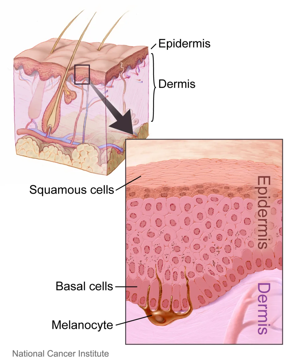
Layers of the skin, with a close-up of the epidermis.
Labeled: epidermis, dermis, squamous cells, basal cells, melanocyte.
Image: Layers of the skin.jpg by Don Bliss (illustrator), National Cancer Institute, Public domain. Resized to 1000 px wide and converted to WebP. Click the image to open it full size.
About these questions
The questions were written with AI in the format PDFStudy produces, then checked question by question against OpenStax Anatomy and Physiology 2e, a free peer-reviewed textbook: wrong or ambiguous items were corrected or removed, and repeats were dropped. Last reviewed 2026-10-20. Found a mistake? Tell us and we will fix it.
Studying from your own lecture slides or textbook? Upload the PDF and PDFStudy turns it into a quiz like this one, which you can also export to Anki.