Heart Anatomy Quiz
94 multiple-choice questions with answers and explanations
Test yourself on the heart: its position and coverings, the four chambers and great vessels, the valves and the path of blood, the coronary arteries, the conduction system, the cardiac cycle and heart sounds, cardiac output, and how the heart develops.
Read a question, pick an option, then open the answer to check yourself and read why it is correct.
Heart position and coverings
Question 1
Which superior surface of the heart is attached to the great vessels?
Show answer
Answer: D. Base
The base is the broad superior surface where the great vessels attach.
Question 2
Which description identifies the apex of the heart?
Show answer
Answer: C. The inferior tip directed slightly to the left of the sternum
The apex is the tapering inferior tip of the heart.
Question 3
The anterior surface of the heart lies deep to which structures?
Show answer
Answer: C. Sternum and costal cartilages
The anterior surface of the heart lies deep to the sternum and costal cartilages.
Question 4
What is the main structural role of the fibrous pericardium?
Show answer
Answer: B. Protecting the heart and maintaining its position in the thorax
The tough connective tissue of the fibrous pericardium supports and protects the heart.
Question 5
Which serous pericardial layer is fused to the fibrous pericardium?
Show answer
Answer: B. Parietal pericardium
The parietal layer attaches to the fibrous pericardium. The visceral layer attaches to the heart.
Question 6
Which heart-wall layer is also called the visceral pericardium?
Show answer
Answer: A. Epicardium
The epicardium belongs to the heart wall and forms the visceral layer of the serous pericardium.
Question 7
Why is serous fluid present around the beating heart?
Show answer
Answer: A. To reduce friction during contraction
Lubricating serous fluid allows the heart to move with less friction.
Question 8
Which sequence lists the heart-wall layers from superficial to deep?
Show answer
Answer: D. Epicardium, myocardium, endocardium
The epicardium is external, the myocardium is in the middle, and the endocardium lines the inside.
Question 9
Which heart-wall layer produces most of the force for pumping blood?
Show answer
Answer: A. Myocardium
The myocardium is the thick muscular layer whose contraction drives blood through the heart.
Question 10
Which layer covers the heart valves and lines the chambers?
Show answer
Answer: D. Endocardium
The endocardium provides the lining exposed to blood within the chambers and over the valves.
Question 11
How does rapidly accumulating pericardial fluid reduce cardiac filling in tamponade?
Show answer
Answer: B. It compresses the heart and prevents full relaxation
Excess pericardial fluid presses on the heart, limiting relaxation and the volume available for the next beat.
Question 12
Which leaf-like extensions near the superior surface of the heart can fill with blood and empty into the atria?
Show answer
Answer: B. Auricles
Auricles are thin-walled atrial appendages that can hold blood and empty into the atria.
Chambers and blood flow
Question 13
Which vessel receives blood directly ejected by the right ventricle?
Show answer
Answer: C. Pulmonary trunk
The right ventricle ejects blood into the pulmonary trunk before it reaches the pulmonary arteries.
Question 14
Where does blood in the pulmonary trunk go immediately after that vessel bifurcates?
Show answer
Answer: D. Left and right pulmonary arteries
The pulmonary trunk divides into pulmonary arteries directed toward the two lungs.
Question 15
Which sequence correctly traces blood returning from systemic capillaries toward the heart?
Show answer
Answer: B. Venules, larger veins, venae cavae
Systemic capillaries drain into venules, which unite into larger veins that ultimately reach the venae cavae.
Question 16
Which partition separates the right and left atria?
Show answer
Answer: D. Interatrial septum
The interatrial septum is the wall between the two atria.
Question 17
What does the adult fossa ovalis mark?
Show answer
Answer: B. The remnant of the fetal foramen ovale
The fossa ovalis is the depression left by the fetal opening in the interatrial septum.
Question 18
Why is the interventricular septum thicker than the interatrial septum?
Show answer
Answer: A. Ventricles generate greater pressure during contraction
The partition between the ventricles must withstand the greater pressures they develop.
Question 19
Which vessel drains blood from the lower limbs and abdominopelvic region into the heart?
Show answer
Answer: C. Inferior vena cava
The inferior vena cava returns blood from regions below the diaphragm.
Question 20
Which muscular ridges are prominent on the anterior internal surface of the right atrium?
Show answer
Answer: C. Pectinate muscles
Pectinate muscles form the ridges of the anterior right atrium and its auricle.
Question 21
Which structures form muscular ridges along the ventricular walls?
Show answer
Answer: A. Trabeculae carneae
Trabeculae carneae are ventricular muscular ridges covered by endocardium.
Question 22
Which chamber contains a moderator band that participates in cardiac conduction?
Show answer
Answer: A. Right ventricle
The moderator band crosses the right ventricle. The left ventricle has no corresponding band.
Question 23
Why does the left ventricle have a thicker muscular wall than the right ventricle?
Show answer
Answer: A. It must overcome greater resistance in the systemic circuit
Both ventricles pump the same volume, but the left needs more pressure to overcome systemic resistance.
Valves and their supports
Question 24
Which valve guards the opening from the right atrium to the right ventricle?
Show answer
Answer: C. Tricuspid valve
The tricuspid valve is the right atrioventricular valve.
Question 25
Which valve lies at the ventricular outlet into the pulmonary trunk?
Show answer
Answer: D. Pulmonary semilunar valve
The pulmonary valve guards the outlet from the right ventricle into the pulmonary trunk.
Question 26
Which valve prevents blood in the aorta from flowing back into the left ventricle?
Show answer
Answer: D. Aortic semilunar valve
The aortic valve closes against blood attempting to return from the aorta.
Question 27
Which strands attach atrioventricular valve flaps to papillary muscles?
Show answer
Answer: B. Chordae tendineae
Chordae tendineae are connective tissue cords linking valve flaps to papillary muscles.
Question 28
What do contracting papillary muscles do during ventricular contraction?
Show answer
Answer: D. Tense the chordae tendineae to prevent valve flaps from turning into the atria
Papillary muscle contraction stabilizes the atrioventricular valve flaps through tension on their cords.
Question 29
Which pair of valves lacks chordae tendineae and papillary muscles?
Show answer
Answer: D. Aortic and pulmonary valves
Both semilunar valves close using their pocket-like cusps rather than tethering cords.
Question 30
During ventricular ejection into the great arteries, which valve arrangement is expected?
Show answer
Answer: B. Atrioventricular valves closed and semilunar valves open
Ejection requires open semilunar valves while closed atrioventricular valves prevent backflow into the atria.
Question 31
What closes a semilunar valve as its ventricle relaxes?
Show answer
Answer: C. Blood moving back toward the ventricle fills its pocket-like cusps
Reverse-directed blood fills the semilunar cusps and brings them together.
Question 32
Which structure provides dense connective tissue rings for heart-valve attachment?
Show answer
Answer: A. Cardiac skeleton
The cardiac skeleton reinforces the valve openings and anchors the valves.
Question 33
A valve cusp is forced backward after its supporting cords are damaged. What is this displacement called?
Show answer
Answer: B. Valve prolapse
Prolapse describes a cusp being pushed backward. Broken chordae can allow this failure of support.
Question 34
What does regurgitation mean in valvular heart disease?
Show answer
Answer: D. Blood flows backward because a valve does not close properly
An incompletely closing valve permits blood to reverse its normal direction.
Question 35
Which valve change in stenosis increases the heart's workload?
Show answer
Answer: B. Rigid valve cusps interfere with forward blood flow
Stenotic valves lose flexibility, making it harder for the heart to propel blood through them.
Coronary circulation
Question 36
When does coronary blood flow reach its peak according to OpenStax?
Show answer
Answer: A. When the heart muscle is relaxed
Coronary circulation cycles with heart activity and peaks during relaxation.
Question 37
From which vessel do the coronary arteries originate?
Show answer
Answer: C. The first portion of the aorta leaving the left ventricle
The coronary arteries branch from the beginning of the aorta to supply the heart itself.
Question 38
Which artery arises from the left coronary artery and follows the coronary sulcus to the left?
Show answer
Answer: B. Circumflex artery
The circumflex is a left coronary branch that follows the coronary sulcus.
Question 39
The left anterior descending artery is another name for which vessel?
Show answer
Answer: D. Anterior interventricular artery
The anterior interventricular artery is also called the LAD and branches from the left coronary artery.
Question 40
What is a vascular anastomosis?
Show answer
Answer: B. An interconnection formed where blood vessels unite
Anastomoses connect vessels and may offer an alternate route around a partial blockage.
Question 41
Why may a coronary blockage cause tissue death despite coronary anastomoses?
Show answer
Answer: D. The heart's anastomoses are very small and provide limited alternate flow
The small size of coronary connections limits their ability to compensate for a blockage.
Question 42
Which regions are supplied by marginal arteries arising from the right coronary artery?
Show answer
Answer: B. Superficial portions of the right ventricle
The right marginal branches supply superficial regions of the right ventricle.
Question 43
In the usual pattern described by OpenStax, which artery gives rise to the posterior interventricular artery?
Show answer
Answer: C. Right coronary artery
OpenStax describes the posterior interventricular artery as a branch of the right coronary artery on the posterior heart surface.
Question 44
Which vein initially parallels the anterior interventricular artery?
Show answer
Answer: D. Great cardiac vein
The great cardiac vein drains the territory supplied by the anterior interventricular artery.
Question 45
Which vein parallels and drains the territory of the posterior interventricular artery?
Show answer
Answer: B. Middle cardiac vein
The middle cardiac vein follows the posterior interventricular artery's territory.
Question 46
How does drainage by the anterior cardiac veins differ from drainage through the coronary sinus?
Show answer
Answer: C. They drain directly into the right atrium, bypassing the coronary sinus
The anterior cardiac veins provide a direct venous route from the anterior right ventricle to the right atrium.
Question 47
What distinguishes myocardial infarction from reduced coronary flow alone?
Show answer
Answer: B. Death of cardiac muscle cells
Myocardial infarction involves death of heart muscle cells after inadequate perfusion and oxygen delivery.
Muscle and electrical conduction
Question 48
What is autorhythmicity in cardiac muscle?
Show answer
Answer: A. The ability to initiate electrical potentials at a regular rate
Autorhythmicity allows cardiac tissue to generate the electrical activity that triggers its contractions.
Question 49
Which cells specialize in initiating and propagating the heart's action potentials?
Show answer
Answer: A. Myocardial conducting cells
Conducting cells are specialized cardiac muscle cells that initiate and distribute electrical impulses.
Question 50
Why is a long refractory period essential in cardiac muscle?
Show answer
Answer: A. It prevents sustained tetanic contraction that would stop effective pumping
The heart needs relaxation between contractions to fill. A long refractory period prevents tetany.
Question 51
Where is the sinoatrial node located?
Show answer
Answer: B. In the superior and posterior right atrial walls near the superior vena cava opening
The SA node lies in the upper posterior right atrium close to the superior vena cava.
Question 52
Why does the SA node normally set the heart's pace?
Show answer
Answer: C. It has the highest inherent rate of depolarization
The SA node reaches threshold faster than the other conducting components, so it normally sets the rhythm.
Question 53
What is the functional importance of the delay at the AV node?
Show answer
Answer: B. It allows atrial contraction to finish before ventricular activation
The AV nodal pause separates atrial and ventricular activation so the atria can finish filling the ventricles.
Question 54
Which sequence correctly follows the impulse after it leaves the AV node?
Show answer
Answer: D. Atrioventricular bundle, bundle branches, Purkinje fibers
The impulse passes down the atrioventricular bundle, through its branches, and into Purkinje fibers.
Question 55
In which direction does ventricular contraction normally spread?
Show answer
Answer: C. From the apex toward the base
Activation begins near the apex and spreads toward the base, helping eject blood into the outflow vessels.
Question 56
Which ion influx is especially important for the plateau of a cardiac contractile cell's action potential?
Show answer
Answer: C. Calcium influx
Calcium entering through slow channels helps maintain the prolonged plateau.
Question 57
Which electrical event does the P wave represent on an ECG?
Show answer
Answer: B. Atrial depolarization
The P wave records atrial depolarization, which precedes atrial contraction.
Question 58
Which ECG feature represents ventricular depolarization?
Show answer
Answer: B. QRS complex
The QRS complex reflects the electrical activation of the ventricles.
Question 59
Why is atrial repolarization usually not seen as a separate wave on a normal ECG?
Show answer
Answer: A. It occurs during the QRS complex and is masked by it
The larger ventricular signal obscures atrial repolarization during the QRS complex.
Cardiac cycle and heart sounds
Question 60
Which term describes the contraction phase of a heart chamber?
Show answer
Answer: B. Systole
Systole is the period when a chamber contracts to propel blood.
Question 61
Which term describes relaxation as heart chambers fill with blood?
Show answer
Answer: B. Diastole
Diastole is the relaxation period that permits cardiac filling.
Question 62
How does most ventricular filling occur in a normal cardiac cycle?
Show answer
Answer: D. Passively through open atrioventricular valves during relaxation
Most filling occurs as blood passes from relaxed atria into relaxed ventricles through open AV valves.
Question 63
What is the atrial kick?
Show answer
Answer: C. The final contribution to ventricular filling produced by atrial contraction
Atrial contraction adds the final portion of blood to the ventricles before ventricular systole.
Question 64
Which volume is measured in a ventricle just before it contracts?
Show answer
Answer: C. End diastolic volume
End diastolic volume is the blood present after filling and immediately before ventricular contraction.
Question 65
Why does ventricular volume remain constant during isovolumic contraction?
Show answer
Answer: A. The atrioventricular valves have closed and the semilunar valves have not yet opened
Early ventricular contraction raises pressure while both inlet and outlet valves are closed, so blood volume stays constant.
Question 66
What pressure relationship begins ventricular ejection?
Show answer
Answer: C. Ventricular pressure exceeds pressure in the aorta or pulmonary trunk
The outflow valves open when ventricular pressure becomes high enough to overcome arterial pressure.
Question 67
What is end systolic volume?
Show answer
Answer: D. Blood remaining in a ventricle after contraction
The ventricle does not empty completely during systole. The residual blood is its end systolic volume.
Question 68
Which valve state characterizes isovolumic ventricular relaxation?
Show answer
Answer: D. Both atrioventricular and semilunar valves are closed
Semilunar valves close before the AV valves reopen, leaving ventricular volume unchanged during early relaxation.
Question 69
What allows the atrioventricular valves to reopen during late ventricular diastole?
Show answer
Answer: D. Ventricular pressure falls below atrial pressure
As ventricular pressure falls below atrial pressure, blood moves into the ventricles and pushes the AV valves open.
Question 70
Which event produces the first normal heart sound, S1?
Show answer
Answer: D. Closure of the atrioventricular valves
S1 accompanies closure of the AV valves at the start of ventricular contraction.
Question 71
Which event produces the second normal heart sound, S2?
Show answer
Answer: C. Closure of the semilunar valves
S2 accompanies aortic and pulmonary valve closure during ventricular diastole.
Cardiac output and control
Question 72
What does cardiac output measure?
Show answer
Answer: B. Blood pumped by each ventricle in one minute
Cardiac output is a flow per minute for each ventricle, rather than the combined output of both ventricles.
Question 73
If heart rate is 80 beats per minute and stroke volume is 70 mL per beat, what is cardiac output for one ventricle?
Show answer
Answer: A. 5.6 L/min
Multiply 80 by 70 to obtain 5600 mL/min, or 5.6 L/min.
Question 74
A ventricle has an end diastolic volume of 120 mL and an end systolic volume of 50 mL. What is its stroke volume?
Show answer
Answer: D. 70 mL
Stroke volume is the difference between the filled ventricular volume and the volume left after contraction.
Question 75
Which calculation gives ejection fraction?
Show answer
Answer: D. Stroke volume divided by end diastolic volume
Ejection fraction expresses the proportion of the filled ventricular volume ejected in one beat.
Question 76
What is cardiac reserve?
Show answer
Answer: A. The difference between maximum and resting cardiac output
Cardiac reserve describes how much cardiac output can increase above its resting value.
Question 77
Which change can make cardiac output fall when heart rate becomes very high?
Show answer
Answer: B. Shorter diastole reduces ventricular filling and stroke volume
At very high rates, reduced diastolic filling can lower stroke volume so much that output falls.
Question 78
Which nerve carries parasympathetic signals that slow the heart?
Show answer
Answer: C. Vagus nerve
The vagus carries cardioinhibitory parasympathetic impulses to the heart.
Question 79
Which neurotransmitter released by sympathetic cardiac nerves increases heart rate?
Show answer
Answer: B. Norepinephrine
Sympathetic cardiac nerves release norepinephrine, which accelerates cardiac electrical activity.
Question 80
What cardiac response helps compensate for increased arterial pressure and baroreceptor stretch?
Show answer
Answer: A. Decreased sympathetic and increased parasympathetic stimulation
Increased stretch signals the cardiac centers to reduce sympathetic drive and increase parasympathetic drive.
Question 81
Within physiological limits, how does greater ventricular filling affect contraction force and stroke volume through the Frank-Starling mechanism?
Show answer
Answer: A. Contraction force increases and stroke volume increases
Within physiological limits, greater initial ventricular muscle-fiber length produces a stronger contraction and increases stroke volume.
Question 82
With end diastolic volume held constant, what does a more forceful ventricular contraction generally do?
Show answer
Answer: A. Increases stroke volume and decreases end systolic volume
A stronger contraction ejects more of the starting blood and leaves a smaller residual volume.
Heart development
Question 83
Which tissue gives rise to the heart during development?
Show answer
Answer: C. Mesoderm
The embryonic heart develops from mesoderm.
Question 84
Where is the cardiogenic area in which heart development begins?
Show answer
Answer: A. Near the head of the embryo
The early cardiogenic area lies near the embryonic head.
Question 85
What change turns cardiogenic cords into endocardial tubes?
Show answer
Answer: D. A lumen develops within each cord
A lumen forms within each cardiogenic cord, converting it into an endocardial tube.
Question 86
What forms when the two endocardial tubes fuse?
Show answer
Answer: C. A single primitive heart tube
The two endocardial tubes come together to create one primitive heart tube.
Question 87
Which sequence lists primitive heart-tube regions from head to tail?
Show answer
Answer: A. Truncus arteriosus, bulbus cordis, primitive ventricle, primitive atrium, sinus venosus
The primitive tube has five regions arranged in this head-to-tail order.
Question 88
Which primitive heart region initially receives all venous blood?
Show answer
Answer: C. Sinus venosus
Early venous inflow enters the sinus venosus before contractions move blood toward the head end of the tube.
Question 89
Which adult vessels develop from the truncus arteriosus?
Show answer
Answer: D. Ascending aorta and pulmonary trunk
The truncus arteriosus divides into the two major ventricular outflow vessels.
Question 90
Which adult chamber develops from the bulbus cordis?
Show answer
Answer: C. Right ventricle
The bulbus cordis gives rise to the right ventricle.
Question 91
Which adult chamber develops from the primitive ventricle?
Show answer
Answer: C. Left ventricle
Despite its general name, the primitive ventricle specifically forms the adult left ventricle.
Question 92
Which adult structures develop from the primitive atrium?
Show answer
Answer: A. Anterior portions of both atria and both auricles
The primitive atrium contributes to the front portions of the two atria and their auricles.
Question 93
Which group of adult structures develops from the sinus venosus?
Show answer
Answer: A. Posterior right atrium, SA node, and coronary sinus
The sinus venosus contributes to the posterior right atrium, its pacemaking node, and the coronary sinus.
Question 94
What is the result of S-shaped folding of the primitive heart tube?
Show answer
Answer: D. Chambers and major vessels move into an alignment resembling the adult heart
Folding repositions the developing chambers and vessels into a layout closer to the adult arrangement.
Heart Anatomy labeled diagrams
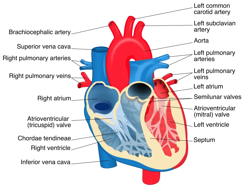
Inside the heart: chambers, valves and great vessels.
Labeled: brachiocephalic artery, left common carotid artery, left subclavian artery, aorta, superior vena cava, inferior vena cava, right pulmonary arteries, left pulmonary arteries, right pulmonary veins, left pulmonary veins, right atrium, left atrium, atrioventricular (tricuspid) valve, atrioventricular (mitral) valve, semilunar valves, chordae tendineae, right ventricle, left ventricle, septum.
Image: Heart diagram (en).svg by ZooFari, text added by Rehua, CC BY-SA 3.0. Rendered from SVG on a white background at 1000 px wide and converted to WebP. Click the image to open it full size.
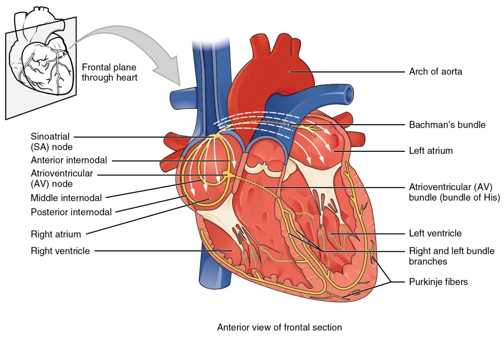
The conduction system of the heart, anterior view of a frontal section.
Labeled: sinoatrial (SA) node, anterior, middle and posterior internodal pathways, Bachmann’s bundle, atrioventricular (AV) node, atrioventricular (AV) bundle (bundle of His), right and left bundle branches, Purkinje fibers, right atrium, left atrium, right ventricle, left ventricle, arch of aorta.
Image: 2018 Conduction System of Heart.jpg by OpenStax College, CC BY 3.0. Resized to 1000 px wide and converted to WebP. Click the image to open it full size.
About these questions
The questions were written with AI in the format PDFStudy produces, then checked question by question against OpenStax Anatomy and Physiology 2e, a free peer-reviewed textbook: wrong or ambiguous items were corrected or removed, and repeats were dropped. Last reviewed 2026-10-20. Found a mistake? Tell us and we will fix it.
Studying from your own lecture slides or textbook? Upload the PDF and PDFStudy turns it into a quiz like this one, which you can also export to Anki.